
Research Article
Received: 2026-04-09 | Revised:2026-05-03 | Accepted: 2026-05-04 | Published: 2026-06-04
Pages: 35-48
DOI: https://doi.org/10.56717/jpp.2026.v05i01.051
Abstract
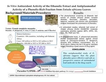
Growth inhibition assays of Plasmodium parasites have become an important strategy for antimalarial drug discovery. Entada africana is a prominent medicinal plant greatly utilized in traditional treatment of varieties of diseases. In this study, the ethanolic extract and phenolic-rich fraction of E. africana leaves were prepared. Following the phytochemical analysis, the ethanolic extract was investigated for its in vitro antioxidant activities, with investigation involving parameters such as ABTS, DPPH, OH, H2O2, NO, FRAP and TAC. An in vitro antimalarial study was carried out on phenolic-rich fraction, using a Plasmodium growth inhibition assay against a clinical isolate of Plasmodium falciparum, cultured in RPMI 1640 growth medium. The results revealed the presence of alkaloids, flavonoids, phenolics, glycosides, saponins, terpenes and tannins with phenolics constituting the greatest portion in the quantitative analysis. In vitro antioxidant assays demonstrated that the extract elicited promising free radical scavenging activities, which could be due to its prominent phenolic components. The growth inhibition assay exhibited good in vitro antiplasmodial, properties with a 50% inhibitory concentration, (IC50) of 28.45 µg/mL, indicating that the phenolic-rich fraction of E. africana exhibited moderate activity against chloroquine-sensitive P. falciparum. These results provide justification for the correlation between oxidative stress management by antioxidants and antimalarial properties of the extract, hence the traditional use of E. africana for the treatment of malaria and the basis for further studies to characterize the extract.
Abstract Keywords
E. africana, leaves, phenolic-rich fraction, in vitro antioxidant activity, antiplasmodial activity, Plasmodium falciparum.
1. Introduction
Malaria poses a significant threat to human health and is the primary cause of death and morbidity in geographical areas where it is endemic [1]. About 92% of children under the age of 5 years and pregnant women who are deficient in immunity are greatly affected by malaria [2]. The most affected regions include sub-Saharan Africa, Asia, Central America, and Latin America, despite the diverse distribution of the disease. Approximately 50% of the global population resides in regions where malaria is a major concern [3]. The most prominent tropical diseases are caused by Plasmodium falciparum, whereas, while the disease caused by Plasmodium vivax, Plasmodium ovale and Plasmodium malariae is generally milder and rarely fatal [4]. The fifth type, Plasmodium knowlesi, rarely causes malaria in humans but is largely responsible for malaria in macaques. This species is currently known as a zoonosis, causing a zoonotic malaria that is majorly found in Southeast Asia, especially in Malaysia [5]. Plasmodium parasites grow and proliferate in cycles in both humans and female Anopheles mosquito vectors [6]. Moreover, P. falciparum has been linked to almost 95% of deaths in sub-Saharan Africa [7].
Globally, the number of malaria cases in 2022 was estimated to be 249 million, which was significantly higher than that in 2019, before the start of the COVID-19 pandemic [7]. According to the WHO [8], the number of malaria cases in 2023 increased to 263 million. Based on this estimation, an incidence of 60.4 cases per 1000 population at risk was reported, and the number of deaths was estimated to 597 000, with a mortality rate of 13.7 per 100 000. The WHO [8] reported that the African region carries the heaviest malaria burden, accounting for 94% and 95% of estimated malaria cases and deaths worldwide, respectively, in 2023. The top five countries carrying the heaviest estimated burden of malaria cases were Nigeria (26%), the Democratic Republic of the Congo (13%), Uganda (5%), Ethiopia (4%) and Mozambique (4%) [8]. Hence, to combat malaria, there is still a necessity to advocate for the control of mosquito vectors, provide treated nets, find new antimalarial compounds, and develop effective vaccines.
However, the development of an effective vaccine has proven to be very difficult [9]. Despite the substantial progress in the treatment of parasitic diseases, malaria remains a significant endemic disease due to factors such as the widespread resistance of malaria parasites to currently available anti-malarial agents, the resistance of mosquito vectors to currently available insecticides, the limited success in the development of malarial vaccines, and the debilitating adverse reactions of orthodox anti-malarial drugs [6], coupled with their high costs [10]. However, traditional medicine practitioners (TMPs) have been using plants from various botanical sources to treat and cure malaria for decades [11]. These medicinal plants have made and continue to make significant contributions to malaria management as they contain molecules with a wide variety of structures and biological activities [12]. Therefore, ethnobotanical investigations of traditional medicines may provide important sources of new antimalarial compounds.
Although in vivo models using Plasmodium berghei provide valuable insights into host–parasite interactions and disease modulation, they do not fully reflect the biology of human malaria [13]. Following our previous in vivo evaluation of the antioxidant effects of Entada africana in P. berghei infected mice, the current study investigated its direct antioxidant and antiplasmodial activities in vitro against chloroquine-sensitive Plasmodium falciparum. This study applied an approach in concordance with bioassay-guided fractionation, where a certain fraction is selected based on phytochemical abundance and pharmacological plausibility [14]. This complementary approach revealed the mechanistic activity of E. africana extract against the human malaria parasite and strengthened the translational relevance of the findings.
The general objective of this study was to carry out in vitro investigation of the antimalarial potential of the leaf extract of E. africana, evaluating its antioxidant and antiplasmodial activities against a clinical isolate of P. falciparum, as a substantial step in the development of a new antimalarial.
2. Materials and methods
2.1. Sample collection and authentication
The leaves of E. africana were collected from the Agba Dam area of Ilorin, Kwara State, Nigeria. The area is located between latitude 8°28′ 33′′N and longitude 4°35′ 22′′E. They were transported in perforated bags to the laboratory of the Plant Biology Department, University of Ilorin, where verification and authentication was done by a taxonomist. The Voucher number (UILH/004/960) was assigned and the specimen was deposited in the Herbarium.
2.2. Plasmodium falciparum
The clinical isolate of P. falciparum was obtained from infected blood samples from malaria patients at the Gitwe District Hospital, Southern Province, Ruhango District, Republic of Rwanda.
2.3. Preparation of plant extract
The plant extract was prepared in accordance with the method described by Adamu et al., [15]. Fresh Entada africana leaves were obtained, well washed, air-dried and pulverized into powder. The powdered plant (700 g) was percolated in 7.0 L of ethanol in a flask, plugged with cotton wool. The mixture was kept on an orbital shaker at 190-220 rpm for 24 h to optimize the extraction process. After extraction, the ethanol extract was filtered using a clean muslin cloth and a Whatman No. 1 filter paper to remove the residues. The filtrate was concentrated at 50 °C under vacuum in a rotary evaporator (RE- 52A, Searchtech Instruments) and then dried at 45 °C in oven to remove the ethanol. The extract obtained was weighed and the percentage yield was calculated using the following formula. The final extract was stored in an airtight sterile bottle at 4 °C inside the refrigerator for the determination of various parameters in the study.

2.4. Determination of phytochemicals
Phytochemical analysis was carried out on ethanolic leaf extract of E. africana (ELEA), both qualitatively [16] and quantitatively for alkaloids [17], total phenolics [18], total tannins concentration [19] and total flavonoid [20].
2.4.1. Quantitative phytochemical analysis of E. africana leaves
2.4.1.1. Determination of total alkaloid concentration
The total alkaloid concentration was estimated using the UV Spectrophotometry procedures described by Ajanal et al. [17]. Briefly, 10 % dimethylsulfoxide (DMSO) was used to dissolve 1 mg of ELEA. The resulting mixture was then added to 1 mL of 2 N HCl and filtered. Then, 5 mL of each of phosphate buffer and bromocresol green were added, and the mixture was transferred to a separating funnel. With vigorous shaking, the mixture was fractionated with 1, 2, 3, and 4 mL of chloroform to remove the complex formed. The fraction was collected in a 10 mL volumetric flask and chloroform was added to dilute it. The absorbance of the complex was read at 470 nm. The total alkaloids in the E. africana leaf extract was represented as mg/100 g of sample as the average of three estimations.
2.4.1.2. Determination of total phenolic concentration
The total phenolic content of E. africana leaf extract was carried out according to the Folin – Ciocalteu reagent procedure described by Tambe and Bhambar [18]. Briefly, 1 g of dried ethanolic leaf extract of E. africana (ELEA) was re-dissolved in 25 mL of ethanol and filtered using Whatmann No. 1 filter paper. In a 25 mL volumetric flask, 1 mL of the filtered extract was mixed together with 9 mL of distilled water. Folin-Ciocalteu phenol reagent was added to the mixture and allowed to react for 5 min, followed by addition of 10 mL of 7 % Na2CO3 w/v solution. The mixture was made to 25 mL with distilled water and incubated for 90 min at room temperature before measuring the absorbance at 750 nm. A standard curve of gallic acid was prepared after calibrating with the spectrophotometer. The estimated total phenolic concentration in E. africana leaf extract was represented as mg of gallic acid equivalent per 100 g of sample.
2.4.1.3. Determination of total tannin concentration
The total tannins concentration was estimated according to the UV Spectrophotometry procedures described by Ekwueme et al. [19]. Briefly, ELEA (1 g) was marcerated with 50 mL of methanol and the resulting mixture was filtered. An aliquot (0.3 mL) of 0.1 N ferric chloride in 0.1 N HCl and 0.3 mL of 0.0008 M potassium ferricyanide were added to 5 mL of the filtrate. The standard curve of gallic acid was prepared after calibrating with the spectrophotometer. The absorbance of the mixture was measured at 720 nm. The total tannin concentration in the E. africana leaf extract was estimated as mg of gallic acid equivalent per 100 g of the sample.
2.4.1.4. Determination of total flavonoid concentration
The total flavonoid content in the E. africana leaf extract was estimated using a modified colorimetric procedure, as outlined by Vabkova et al. [20]. Briefly, a sufficient quantity (1 g) of ELEA was dissolved in 5 mL of ethanol and filtered. In a test tube, 0.5 mL of filtered ELEA and 0.5 mL of distilled water were added. Following the addition of 0.3 mL of 10% AlCl3, and 0.3 mL of 5% NaNO2 were added to the mixture. After 5 min, 2 mL of 1 M NaOH was added. At 15 min later, the absorbance was read at 510 nm in comparison to a blank. Different catechin concentrations were used to generate standard curves. Catechin equivalents (CE) per 100 g of dry weight was used to express the flavonoid content in mg.
2.5. Antioxidant study
2.5.1 Determination of 2,2-azinobis-(3-ethylbenzothiazoline-6-sulfonic acid) (ABTS) radical scavenging activity
The ABTS radical scavenging activity of extract and the standard were determined in accordance with the method described by Re et al. [21], with slight. modification. Briefly, the stock solution of ABTS was diluted with 70% ethanol to an absorbance of 0.75 ± 0.05, to obtain working solution. ABTS radical cations (ABTS+) were generated by reacting an ABTS stock solution (7 mM) with 7 mM potassium persulphate in a ratio 2:1 and the mixture was allowed to stand in the dark at room temperature for 16 h before use. Butylated hydroxyl toluene (BHT) was used as the reference antioxidant solution. Then 2 mL of ABTS working solution was mixed with 50 µL of different concentration (10, 20, 30 and 40 µg/mL) of the extract/BHT and the absorbances were measured after 20 min at 734 nm. The percentage inhibition of each mixture was determined using the formula described by Guchu et al. [22].

Where, Ac and As represent the absorbance of the blank and sample, respectively.
2.5.2. Determination of 2,2-diphenyl-1-picrylhydrazyl (DPPH) radical scavenging activity
The DPPH radical scavenging activity of the extract was determined using 2,2-diphenyl-1-picrylhydrazyl (DPPH) radical in accordance with the procedures described by Hwang et al. [23] with minor modifications. Briefly, the extract was prepared in ethanol (analytical grade) at 4 different concentrations (10, 20, 30 and 40 µg/mL). Then 10 µL of each of the extract concentrations was added to an equal volume of 100 µM DPPH reagent (10 µL) in ethanol solution in a well-labelled, clean 96-well microtitre plate (Tarsons, Kolkata, India). Standard solutions of the antioxidant butylated hydroxyl toluene (BHT) and reagent blank (10 µL each) were treated similarly. All mixtures were shaken well and allowed to stand for 30 min. After the incubation period, each mixture was measured against a standard, butylated hydroxyl toluene (BHT) for absorbance at 517 nm using an ELISA reader specified for microtitre plates (Bio Rad Laboratories Inc, California, USA, Model 550). These experiments were performed in triplicate. The percentage inhibition of each mixture was determined using the following formula.
Where, Ac and As represent the absorbances of the blank and sample, respectively.
2.5.3. Determination of hydroxyl radical scavenging activity
The hydroxyl radical scavenging activity of the plant extract was measured according to the method of Halliwell et al. [24]. This determined the interactions between deoxyribose and test extracts for hydroxyl radicals, which were obtained by Fenton’s reaction. Briefly, the reaction mixture contained 1.0 mL of reagent (3.0 mM deoxyribose, 0.1 mM EDTA, 2 mM H2O2, 0.1 mM L-ascorbic acid, 0.1 mM FeCl3.6H2O in 10 mM phosphate buffer, pH 7.4) and 1.0 mL of the extract (50 mg/mL). The reaction mixtures were incubated at 37 °C for 1 h, followed by the addition of 1.0 mL of 1% (w/v) TBA (in 0.25 M HCl) and 1.0 mL 10% (w/v) trichloroacetic acid (TCA). The reaction mixture was heated in a boiling water bath at 100 °C for 20 min, and the pink chromogen (malondialdehyde-(TBA) adduct) was extracted into 1.0 mL of butan-1-ol and the absorbance (Abs) was read at 532 nm against a reagent blank. BHT (butylated hydroxytoluene) was used as a positive control. This experiment was performed in triplicate. The percentage inhibition was calculated using the expression below, which was plotted against concentration.
Where, Ac and As represent the absorbances of the blank and sample, respectively.
Where, Acontrol = absorbance of control (containing all reagents except the test compound) and Asample = absorbance of samples (containing all reagents including the test compound).
2.5.4. Determination of hydrogen peroxide scavenging activity
The ability of the extracts to scavenge hydrogen peroxide was determined according to the method of Hussen and Endalew, [25] with a slight modification. A solution of hydrogen peroxide (2 mM) was prepared in 0.2 M phosphate buffer (pH 7.4). Briefly, about 0.2 M potassium dihydrogen phosphate and 0.2 M sodium hydroxide solutions were prepared. A 50 mL potassium dihydrogen phosphate solution was placed in a 200 mL volumetric flask and 39.1 mL of 0.2 M sodium hydroxide solution was added. Finally, the volume was made up to 200 mL with distilled water to prepare the phosphate buffer (pH-7.4). 50 mL of phosphate buffer solution was added to an equal amount of hydrogen peroxide to generate free radicals and the solution was kept at room temperature for 5 min to complete the reaction. 100 μL of the extract (Conc. 50, 100, 150, 200, 250, and 300 μg/mL in distilled water) was added to 600 μL of hydrogen peroxide solution. After 30 min incubation in the dark, the absorbance at 230 nm was recorded using a UV–Vis Spectrophotometer. A reference stock solution of gallic acid of equivalent concentration was used as the standard solution. Ethanol was used as the blank for the determinations, which were performed in triplicate. The percentage inhibition of each mixture was calculated using the following equation:
Where, Ac and As represent the absorbances of the blank and sample, respectively.
2.5.5. Determination of nitric oxide radical scavenging activity
The scavenging capacity of the extract against the nitric oxide (NO) was evaluated in accordance with the procedures described by [26]. Briefly, the reaction mixture (6 mL), was prepared to include 1 mL of extract (50 mg/mL), 1 mL of phosphate buffer saline (0.01 M) at pH 7.4, and 4 mL of sodium nitroprusside (10 mM). The reaction mixture was incubated at 25 °C for 150 min and an aliquot (0.5 mL) was taken out, then 1 mL of sulphanilic acid reagent (0.33% in 20% glacial acetic acid) was added, then left for 5 min to complete the diazotization reaction. Following this reaction, 1 mL of 0.1% N-(1-naphthyl) ethylenediamine dihydrochloride (NEDD) was added, mixed, and left to stand for additional 30 min under diffused light. Using a UV-Vis spectrophotometer, the absorbances were read at 540 nm against the blank. Gallic acid (50 mg/mL) was used as the standard for comparative antioxidant analysis, and the experiments were performed in triplicate. The percentage inhibition was determined using the following formula:
Where, Ac and As represent the absorbances of the blank and sample, respectively.
2.5.6. Determination of reducing power
The reducing powers of the extract and standard control were determined as ferric reducing antioxidant power (FRAP) in accordance with the method described by Thasneem et al. [27] with slight modifications. Briefly, 1 mL of the test extract (50 mg/mL) was mixed with sodium phosphate buffer (2.5 mL; 0.2 M; pH 6.6) and potassium ferricyanide (2.5 mL; 1%), then incubated (50 °C; 20 min). Trichloroacetic acid (2.5 mL; 10%) was added to the incubated mixture and centrifuged (3000 rpm; 10 min; 4 °C). The supernatant was collected. The supernatant solution (2.5 mL) was mixed with an equal volume of distilled water and fresh ferric chloride solution (0.5 mL; 0.1%) and kept for 10 min. The absorbance of reaction mixture was measured at 700 nm. Gallic acid (50 mg/mL) was used as the standard for comparative antioxidant analysis, and the experiments were performed in triplicate. The following formula below was used to calculate the percentage of inhibition:
Where, Ac and As represent the absorbances of the blank and sample, respectively.
2.5.7. Determination of total antioxidant capacity
The total antioxidant capacity of the extract was determined according to the method described by Prieto et al. [28]. The assay is based on the reduction of Mo(VI) to Mo(V) by the samples and the formation of a green colored phosphate/ Mo(V) complex at acidic pH. Briefly, 0.5 mL (50 mg/mL) of the extract or standard solution was added to 3 mL of the reagent solution which consisted of 0.6 M sulphuric acid, 28 mM sodium phosphate and 4 mM ammonium molybdate. The test tubes containing the reaction mixture were incubated in a water bath at 95 °C for 10 min. The mixture was then allowed to stand and cool to room temperature. The absorbance of the sample was measured at 695 nm against a blank, using a UV-Vis spectrophotometer. Gallic acid (50 mg/mL) was used as the standard for comparative antioxidant activity, that were determined in triplicate. The percentage inhibition was determined using the following formula below:
Where, Ac and As represent the absorbances of the blank and sample, respectively.
2.6 Antiplasmodial study
2.6.1 Extraction and liquid–liquid partitioning of total phenolics from E. africana leaves
Total phenolics were extracted from E. africana leaves following the method described by Al-Farsi and Lee [29], with slight modifications. Briefly, 50 g of powdered leaves were defatted with 125 mL of n-hexane at 60 °C for 4 h and filtered. The defatted residue was air-dried and extracted with 50% acetone (3 L) using a solvent-to-solid ratio of 60:1 (v/w).
The mixture was stirred at 45 °C for 1 h and then mechanically shaken using an orbital shaker at 150 rpm for 4 h daily over a period of 48 h. The extract was filtered, and the extraction process was repeated to ensure maximum recovery.
The combined filtrates were then concentrated at 60 °C. The concentrated extract was subjected to liquid–liquid partitioning with n-butanol. The aqueous layer was separated, and the butanol fraction was collected and concentrated at 60 °C. The final concentrate was then subjected to phenolic test. The resulting phenolic-rich fraction was evaporated to dryness under reduced pressure using a rotary evaporator to obtain a crude total phenolic fraction.
2.6.2. Cultivation of chloroquine sensitive Plasmodium falciparum
P. falciparum parasites were cultured from the clinical samples using the procedures described by Trager and Jensen [30], slight. modifications. Briefly, chloroquine sensitive P. falciparum-infected blood was ascertained using the procedure of Prasad et al. [31], and same was obtained in an EDTA bottle at Gitwe District Hospital, Rwanda. The anticoagulated blood sample was spun at 2000 rpm for 5 min and then aspirated the plasma as well as buffy coat. The red cells obtained were washed three times with 100% washing medium (RPMI 1640 incomplete culture medium). The cells were re-suspended at 5% haematocrit (hct) in complete culture medium (RPMI 1640, supplemented with human plasma from O positive blood). Complete malarial culture medium cMCM (8 mL RPMI + 1 mL Plasma) was pipetted into tissue culture flask with 1 mL red cells (at approx. 1000 parasites/µL). The tissue culture flasks, with caps were loosely screwed, then horizontally placed in a CO2 incubator (JP-Selecta S.A. 4002628) at 37 °C for 4 days. Giemsa staining of the smear was carried out to observe the growth of the P. falciparum isolate.
2.6.3. Evaluation of antiplasmodial activities of phenolic-rich fraction of E. africana leaf
An in vitro antimalarial study was carried out using the method described by Ntalani et al. [32], with slight modifications. Briefly, dried plant extract was prepared at concentrations ranging from 180.00 to 20.00 µg/mL in methanol serial dilution and then 50 µL/well was preloaded in triplicate into a 96-well microtitre plate (9 rows) thereafter 50 µL of Plasmodium culture was added to each well. The negative control well (10th row) contained only the plasmodium culture. The microtitre plate was tightly covered and placed on an orbital rotator shaker for few minutes. The multiwell plate was then incubated in a CO2 incubator for 48 h. Thick drops were then prepared from each well. Thick droplet slides were read using an optical microscope to determine parasitaemia and the parasite growth inhibition percentage was calculated using the following formula. IC50 values were determined using method described by Eseyin et al. [33].

Where, AV. CPM is the average count per minute.
2.7. Data analysis
Data are expressed as the mean ± SEM of at least three determinations. Graphpad prism version 7.0 was used for statistical analyses while statistical significance was set at p < 0.05.
3. Results and discussion
Medicinal plant research has grown over time as a means of identifying promising plants and novel drug candidates to control diseases such as malaria [34]. Compounds found in the extracts of these plants have been explored for their biological activities, as evidenced by in vitro and in vivo investigations of many diseases. Traditional practitioners, as well as various ethnic groups have always found solace in traditional medicines, using various plant parts. In this study, the leaf extract of E. africana was evaluated, and its ethanolic form yielded 17.51% of the powdered sample. A preliminary investigation via phytochemical analyses of the extract was conducted, revealing seven considerable secondary metabolites of varied quantities, as presented in Table 1.
Table 1 Quantitative phytochemical analysis of ethanolic leaf extract of Entada africana.
S/N | Phytochemicals | Concentration (mg/100g) |
1 | Saponins | 0.72 ± 0.01 |
2 | Flavonoids | 9.86 ± 0.64 |
3 | Tannins | 37.40 ± 1.59 |
4 | Alkaloids | 0.70 ± 0.00 |
5 | Glycosides | 0.33 ± 0.00 |
6 | Phenolics | 91.75 ± 0.08 |
7 | Terpenoids | 1.06 ± 0.00 |
Values are expressed as Mean ± SEM. for n=3; SEM.–Standard Error of Mean; n- number of replicates. | ||
For many years, the schizont maturation inhibition assay has been a standard in vitro method for the initial antiplasmodial screening of substances from conventional databases and plants, such as Calotropis procera (Ait). R.Br (Asclepiadaceae), Hedyotis corymbosa Linn. (Rubiaceae), Andrographis paniculata Nees (Acanthaceae), etc. [35].
This study examined the antioxidant and antiplasmodial properties of E. africana leaf extracts in vitro. The investigation mimicked the traditional method of preparation by selecting ethanol as the extractant for E. africana leaves. Following preliminary screening, fractionation was guided by quantitative phytochemical analysis, which revealed that phenolic compounds were the most abundant constituents. The phenolic-rich fraction was prioritized for antiplasmodial evaluation based on its phytochemical predominance and the documented relevance of phenolic compounds to antiplasmodial activity [36]. This method is consistent with bioassay-guided fractionation, which selects fractions according to pharmacological plausibility and phytochemical abundance [14]. Several studies have demonstrated that polyphenolic chemicals, which are commonly present in naturally occurring products, have important biological functions [37]. In this study, only the antioxidant activity of the crude ethanolic extract was reported, as it represents the initial extract under investigation. Antioxidant property of the phenolic-rich fraction was internally used to prioritize the most abundant phytochemicals for downstream in vitro antiplasmodial assays.
The in vitro antioxidant activities were investigated, and the dose-dependent patterns of ABTS and DPPH radical scavenging activities of E. africana leaf extract are presented in Table 2. The stable radical cations were produced when ABTS was oxidized with potassium persulfate, which in the presence of the extract and BTH as hydrogen donating antioxidants, the blue ABTS radical cations were reduced and eventually became decolorized in a dose-dependent pattern. As shown in the table the ABTS scavenging activity increased with increasing concentration of the extract and the reference antioxidant solution (BTH). BTH exhibited antioxidant scavenging activity that was almost two-fold of the value of the plant extract at a tested concentration of 40 μg/mL.
Table 2. Percentage inhibition of ABTS and DPPH radicals by ethanolic leaf extract of Entada africana.
Concentrations (µg/mL)
Inhibition (%) of ABTS
Inhibition (%) of DPPH
Sample
Reference (BHT)
Sample
Reference (BHT)
10
31.02 ± 3.84
44.96 ± 0.51
32.68 ± 2.11
48.55 ± 1.08
20
33.18 ± 4.70
49.64 ± 2.77
38.41 ± 0.94
62.38 ± 0.40
30
39.71 ± 2.05
65.88 ± 1.90
46.66 ± 3.40
87.47 ± 2.13
40
39.92 ± 2.66
66.47 ± 0.18
48.28 ± 1.23
91.34 ± 1.55
Data are expressed as mean ± SD (n = 3, p < 0.05).
In the DPPH assay, the interaction of ELEA or the standard antioxidant compound, BHT, with DPPH is based on the transfer of hydrogen atoms or electrons to the DPPH radical, converting it to 2, 2- diphenyl-1- picrylhydrazine. The reduction in DPPH radicals caused discoloration from purple to pale yellow, indicating the scavenging activity. The antioxidant capacity of the tested doses of ELEA was demonstrated at various concentrations (10, 20, 30 and 40 µg/mL) in a dose-dependent manner. The activities within the extracts varied with increasing concentration, with the 40 µg/mL extract demonstrating the highest DPPH scavenging potential. The results showed that the extract at 40 µg/mL exhibited maximum ability of 48.28% against DPPH radicals as compared to 32.68, 38.41, and 46.66% at concentrations of 10, 20 and 30 µg/mL, respectively. This pattern can be interpreted as dose-dependent inhibition of the radicals generated from DPPH at a level of significance p < 0.05 among each of the tested ELEA concentrations, even though significantly lower (p < 0.05) than the values of BHT, which exhibited almost double of its radical-inhibiting capacity, (91.34 ± 1.55) at the 40 µg/mL.
The ability of ELEA to scavenge ABTS and DPPH radicals was lower than that of the reference antioxidant at all tested concentrations. In the ABTS radical scavenging assay, as shown in Table 2, the plant extract exhibited potent activity against ABTS radicals in concentration-dependent pattern but was significantly lower (p < 0.05) than the reference antioxidant. This is in correlation with the study reported by Chintalapani et al. [38] who found that various solvent extracted crude samples of Sesuvium portulacastrum of the whole plant demonstrated lower (p < 0.05) ABTS radical scavenging activities than the standard antioxidant. In the DPPH assay, the ethanolic leaf extract of E. africana was able to neutralize and absorb the free radicals produced by 2,2-diphenyl-1-picrylhydroxyl, stabilizing the free electrons carried by the free radicals. According to Dehpour et al. [39], redox processes involve the release of electrons by radical scavengers to neutralize the radicals generated by DPPH, a stable nitrogen-centered radical, thereby reducing its destructive potential. The study also reported that any compound capable of donating hydrogen atoms or electrons and changing color from violet to yellow could be classified as an antioxidant and thus a radical scavenger. The moderate DPPH radical scavenging activity of E. africana leaf extract (Table 2) appears to be directly related to the phytochemical content of the leaves. Due to its high phenolic content, the ethanolic leaf extract of E. africana demonstrated varying levels of antioxidant capabilities over a range of concentrations in a dose-dependent manner. This demonstration of antioxidant activity is in agreement with the findings of Jorgensen et al. [40], who suggested that the phenolic chemicals and flavonoids present in the ethanolic plant extract had an antioxidative effect on biological systems.
Table 3 shows the antioxidant activities of ELEA in hydroxyl (OH), hydrogen peroxide (H2O2), nitric oxide (NO), total antioxidant capacity (TAC), and ferric reducing antioxidant power (FRAP) assays. The results presented in the table indicated that the extract elicited significantly (p < 0.05) lower activity but a considerable percentage of inhibition at 50 µg/mL of gallic acid. equivalent. Hydroxyl radical scavenging activity was estimated by generating hydroxyl radicals using ascorbic acid–iron EDTA. This radical is the most reactive oxygen-centered species and causes severe damage to adjacent biomolecules. The hydroxyl radical inhibition potential of the extract was significantly (p < 0.05) lower but exhibited a positive correlation with gallic acid equivalence (GAE). Hydrogen peroxide [H2O2], although not a radical, but another reactive oxygen species, was inhibited by the extract, which demonstrated stronger (p < 0.05) antioxidant activity than gallic acid. Hydroxyl and hydrogen peroxide are examples of reactive oxygen-centered species with the former being the most reactive followed by peroxynitrite (ONOO-) causing indiscriminate damage to adjacent biomolecules such as lipids, proteins and DNA [36]. Erkan et al. [41] suggested that the ability of plant extracts to donate hydrogen or delocalized electrons is indicated by the amount of hydroxyl radical that is mopped up. Several studies have attributed the high concentration of hydrogen peroxide to its capacity to permeate biological membranes which can damage glycolytic enzymes such as glyceraldehyde-3-phosphate dehydrogenase [42]. Ebrahimzadeh et al. [43] suggest that phenolics in extracts could be responsible for H2O2 scavenging, since they could offer it electrons to get neutralized into water and oxygen molecules. The predominant phytochemical group in the E. africana extract (Table 1) may be responsible for the removal of hydrogen peroxide in the assays. Kamalanathan et al. [44] also reported that drugs or agents which quench radicals or reactive oxygen species, serve as better therapeutic for the stress related disorders such as that from malaria. The generation of hydrogen peroxide from free heme, a byproduct of hemoglobin digestion by Plasmodium, was reported to be inhibited by plant extracts which could provide a potent antioxidant-modulating environment, thereby influencing the availability of heme to bind and inhibit glyceraldehyde-3-phosphate dehydrogenase (G3PDH), an important enzyme in its own glycolytic pathway [45]. The inactivation of the parasites’ G3PDH eventually shuts down energy processing and availability for their survival due to the sensitivity to the glycolytic enzyme of the free hemes. This study suggests a possible mechanism of parasite growth inhibition that has been widely reported. Conversely, the integrity of the host’s G3PDH and energy-producing systems in cells may be preserved by consuming plant foods high in antioxidants.
The nitric oxide radical scavenging potential of ELEA was evaluated using 50 mg/mL of the extract and gallic acid at equal concentrations. There was a significant difference (p < 0.05) between the NO radical scavenging potentials of the extract and gallic acid. Nitric oxide is a reactive nitrogen species (RNS), an effector molecule in many biological systems, and a potent pleiotropic modulator of numerous physiological processes [46]. It is also a free radical that can be converted to a stronger oxidant, peroxynitrite, upon reaction with superoxide anion radical (O2*-). Nimse and Pal [47] reported that natural antioxidant compounds of plant origin compete with nitric oxide for superoxide oxygen to prevent the formation of peroxynitrite, which oxidizes a wide range of biomolecules [47]. In this study, ELEA effectively demonstrated NO radical scavenging activity, which corresponds to the study reported by Sasikumar et al. [48] but is significantly lower (p < 0.05) than that of gallic acid, a standard antioxidant. This suggests that the extract contains certain radical-scavenging compounds, providing empirical support for the use of E. africana leaves in the treatment of various diseases.
Ferric reducing antioxidant power (FRAP) is another assay used to evaluate the electron donating ability of E. africana ethanol extract. The results of this study (Table 3) showed that the reducing powers of both ELEA and the reference antioxidant, at 50 mg/mL, indicate that the abilities to reduce Fe3+ to Fe2+ were significantly different (p < 0.05) between the two sources, with a lower activity reported in ELEA. Iron (III) can be reduced to iron (II) in samples by electron donor compounds [49]. FRAP, including other antioxidant parameters of a plant extract, implicates ELEA potential in antimalarial activity. Sadik et al., [50] reported that extracts rich in electron donating compounds play a dual role in in vitro antioxidant and antimalarial activities by scavenging radicals and inhibiting the parasite growth.
The high phenolic content of the E. africana extract may have contributed to its ability to scavenge the free radicals produced by the reagent solutions in the total antioxidant capacity (TAC) experiments (Table 3) at 50 mg/mL. This result supports the claim made by Rice-Evans et al. [51] that plant extracts rich in phenolic compounds could induce redox, singlet oxygen quenching, and hydrogen donating activities.
Table 3. Percentage inhibition of free radicals/reactive oxygen species by ethanolic leaf extract of Entada africana compared with reference antioxidant.
Assays | Inhibition (%) of Free radicals/Reactive oxygen species | ||
ELEA (50 mg/mL) | GAE (50 mg/mL) | ||
Hydroxyl radical scavenging activity | 60.56 ± 0.55 | 85.17 ± 0.08 | |
Hydrogen peroxide scavenging activity | 57.81 ± 1.23 | 36.12 ± 0.25 | |
Nitric oxide inhibitory potential | 15.83 ± 0.61 | 29.05 ± 0.76 | |
Ferric reducing antioxidant power | 13.29 ± 0.92 | 64.52 ± 0.05 | |
Total antioxidant capacity | 12.04 ± 0.08 | 23.19 ± 0.68 | |
Data are expressed as mean ± SD (n = 3, p < 0.05). ELEA- Ethanolic leaf extract of E. africana; GAE- Gallic acid equivalent. | |||
Both the host and parasite are affected by oxidative stress during malarial pathogenesis [52]. The ROS cascade is generated by the activation of neutrophils to facilitate the erythrocytic destruction of the parasite, while ROS are also produced by the parasite via hemoglobin degradation. ROS generated from multiple sources usually result in host tissue damage in areas such as the vascular endothelial lining and blood-brain barrier, undermining the antioxidant defense system. The use of antioxidants strengthens this system and offers protection against the critical stages of malaria [52].
In an antimalarial study, the phenolic-rich fraction of E. africana (6.925% of the powdered sample) was evaluated for its growth-inhibitory activity against a clinical isolate of P. falciparum. The fraction exhibited concentration-dependent growth inhibition of P. falciparum with an IC50 of 28.45 µg/mL (Fig. 1).

Figure 1. In vitro growth inhibition of Plasmodium falciparum by Entada africana phenolics.
Percentage growth inhibition was plotted against log₁₀-transformed compound concentration (µg/mL). Data are presented as mean ± SEM (n = 3). A four-parameter logistic nonlinear regression model was used to determine the IC₅₀ value (28.45 µg/mL).
Phenolics are a major group of compounds that act as primary antioxidants or free radical scavengers [53]. These compounds possess unique features due to their interaction between the π-electrons of the benzene ring and the hydroxyl groups. Most significantly, this connection allows the molecules to produce free radicals that are stabilized by delocalization. Radical-mediated oxidation processes can be altered by the formation of these comparatively long-lived radicals [54]. The reported redox characteristics of phenolic compounds, which can be useful in quenching singlet oxygen, free radicals, or hydrogen peroxides, are primarily responsible for the antioxidant activities of the compounds [53]. The in vitro findings of this study are not in contradiction to those of Builders et al. [55], who reported that the phenolic extract of Parkia biglobosa showed strong antimalarial activity as well as excellent antiplasmodial activity against a clinical isolate of P. falciparum. Saxena et al. [56] reported that phenolics are among the many secondary metabolites of plants that have been shown to suggest a link between antioxidant and antiplasmodial activities. The antiplasmodial activities of many phenolic compounds have also been described by Kim et al. [53]. As reported earlier, the leaf extract of E. africana revealed phenolics (Table 1) as the constituent with the highest quantity which might be responsible for its efficacy as an antimalarial plant concoction and decoction in local settings.
In this study, although the standard drug control, chloroquine phosphate, evaluated against the isolates of P. falciparum gave an IC50 of 0.0061 μg/mL, the IC50 value of E. africana (phenolic-rich fraction) was found to be 28.45 μg/mL (Fig. 1). However, plant extracts have been classified as either good or poor potential drugs based on their IC50 values [57]. More significantly, based on their in vitro antiplasmodial IC50 values, plant extracts' efficacies in terms of whether they include inherent active biological components have been divided into four groups [58]. Based on these standards, the in vitro activity of the phenolic components of E. africana against the clinical isolate of P. falciparum was considered moderately active, with an IC50 of 28.45 μg/mL, falling into the 5-50 μg/mL category, which Kumari et al. [58] reported as good.
The antiplasmodial activity of the phenolic-rich fraction of Entada africana leaves in this study (IC₅₀ = 28.45 µg/mL) exhibited moderate efficacy compared to related species within the same genus, although crude extracts. Notably, ethyl acetate leaf extract of Entada pervillei was reported to exhibit markedly higher potency (IC₅₀ = 0.5 µg/mL), indicating a significantly stronger antiplasmodial effect [59]. In contrast, the activity of various crude extracts of Entada abyssinica leaves and stem bark elicited IC₅₀ ranging between 30.26 and 101.26 µg/mL against chloroquine-sensitive (3D7) and chloroquine-resistant (Dd2) strains [60]. This is comparable to the results of present study, suggesting a similar moderate to weak level of bioactivity between these two species. The marked difference in activity between E. pervillei and the other Entada species may be attributed to solvent variations and phytochemical variability, plant part used, ecological variations, synergistic and antagonistic interactions. Therefore, the phenolic components of E. africana leaves could be considered a good source of lead candidates for the development of antimalarial drugs.
4. Conclusions
Malaria is a devastating tropical disease that poses a financial burden on endemic regions in sub-Saharan Africa. The challenges faced in developing effective antimalarial medications have promoted the use of traditional solutions with medicinal plants. A limited number of these plants have been scientifically explored for their ameliorative activities. The results presented in this study revealed that the ethanolic leaf extract of E. africana has good antioxidant activity. Evidence also suggests that the phenolic constituents of the plant exhibit active antiplasmodial activity, with respect to the magnitude of growth inhibition elicited on a clinical isolate of P. falciparum, although significantly lower than that of chloroquine (reference agent). This in vitro study established antioxidant and antiplasmodial potentials as the dual roles played by the E. africana leaf extract, corresponding to the abundance of phenolic secondary metabolites therein. Therefore, it is essentially to profile the phytoconstituents using chromatographic techniques to aid in the isolation of a potential antiplasmodial lead agent from E. africana leaves.
Disclaimer (artificial intelligence)
The authors hereby state that no generative AI tools, such as Large Language Models (ChatGPT, COPILOT, etc.) and text-to-image generators, were utilized in the preparation or editing of this manuscript.
Authors’ contributions
Conceptualization, B.T.; O.B.; methodology, B.T.; software, B.T..; validation, O.B., S.M.; formal analysis, B.T.; investigation, B.T.; resources, B.T.; data curation, B.T.; writing – original draft preparation, B.T.; writing – review & editing, S.M.; O.B.; visualization, B.T.; supervision, O.B., S.M.; project administration, B.T.; funding acquisition, B.T.
Acknowledgements
The authors are highly grateful to Mr. Bolu Ajayi of Herbarium Section of the Department of Plant Biology and Mr. Dele Aiyepeku of Biochemistry Department Laboratory, University of Ilorin, Nigeria, Rwanda Biomedical Center (RBC), Kigali, Rwanda, Clinical Director, Gitwe District Hospital, Southern Province, Rwanda, Mr. Charles Habimana of Microbiology Department, University of Gitwe, Rwanda and lastly, Central Research and Diagnostic Laboratory, Ilorin, Nigeria, for their various supports during this work.
Funding
This research did not receive any specific grant from funding agencies in the public, commercial, or not-for-profit sectors.
Availability of data and materials
The authors declare that data and materials will be made available by the corresponding author upon request in accordance with the Journal policy.
Conflicts of interest
The authors declare that they have no conflicts of interests.
References
1. | Kwansa-Bentum, B.; Agyeman, K.; Larbi-Akor, J.; Anyigba, C.; Appiah-Opong, R. In vitro assessment of antiplasmodial activity and cytotoxicity of Polyalthia longifolia leaf extracts on Plasmodium falciparum strain NF54. Malaria Res. Treat. 2019, 6976298. https://doi.org/10.1155/2019/6976298 |
2. | Adebayo, J.O.; Krettli, A.U. Potential antimalarials from Nigerian plants: A review. J. Ethnopharmacol. 2011, 133, 289-302. https://doi.org/10.1016/j.jep.2010.11 .024 |
3. | Muluye, A.B.; Melese, E.; Afinew, G.M. Antimalarial activity of 80% methanolic extract of Brassica nigra (L.) Koch. (Brassicaceae) seeds against Plasmodium berghei infection in mice. Complement. Alter. Med. 2015, 15, 367. https://doi.org/10.1186/s12906-015-0893 |
4. | Sutherland, C.J.; Tanomsing, N.; Nolder, D.; Oguike, M.; Jennison, C.; Pukrittayakamee, S.; Dolecek, C.; Hien, T.T.; do Rosário, V.E.; Arez, A.P.; Pinto, J.; Michon, P.; Escalante, A.A.; Nosten, F.; Burke, M.; Lee, R.; Blaze, M.; Otto, T.D.; Barnwell, J.W.; Pain, A.; Williams, J.; White, N.J.; Day, N.P.; Snounou, G.; Lockhart, P.J.; Chiodini, P.L.; Imwong, M.; Polley, S.D. Two nonrecombining sympatric forms of the human malaria parasite Plasmodium ovale occur globally. J. Infect Dis. 2010, 201, 1544-50. https://doi.org/10.1086/652240 |
5. | Singh, B.; Kim Sung, L.; Matusop, A.; Radhakrishnan, A.; Shamsul, S.S.; Cox-Singh, J.; Thomas, A.; Conway, D.J. A large focus of naturally acquired Plasmodium knowlesi infections in human beings. Lancet. 2004, 363, 1017-24. https://doi.org/10.1016/S0140-6736(04)15836-4 |
6. | Centers for Disease Control and Prevention. 2017. https://www.cdc.gov/malaria/about/ biology/ (accessed on June 2017). |
7. | World Health Organization. World malaria report 2023. Geneva: Licence: CC BY-NC-SA 3.0 IGO. 1-5. |
8. | World Health Organization. World malaria report 2024: Addressing inequity in the global malaria response. Geneva, 2024. |
9. | Chattopadhyay, R.; Kumar, S. Malaria vaccine: Latest update and challenges ahead. Indian J. Exp. Biol. 2009, 47, 527-36. |
10. | Enenebeaku, C.K.; Ogukwe, C.E.; Nweke, C.O.; Anyado-Nwadike, S.O.; Obi, M.; Duru, I.A.; Enenebeaku, U.E.; Ogidi, O.I. Antiplasmodial and in vitro antioxidant potentials of crude aqueous and methanol extracts of Chasmanthera dependens (Hochst). Bull. Natl. Res. Cent. 2022, 46, 33. https://doi.org/10.1186/s42269-022-00721-3 |
11. | Asase, A.; Oteng-Yeboah, A.A.; Odamtten, G.T.; Simmonds, M.S. J. Ethnobotanical study of some Ghanaian anti-malarial plants. J. Ethnopharmacol. 2005, 99, 273-279. https://doi.org/10.1016/j.jep.2005.02.020 |
12. | Bero, J.; Frederich, M.; Quetin-Leclercq, J. Antimalarial compounds isolated from plants used in traditional medicine. J. Pharm. Pharmacol. 2009, 61, 1401–33. https://doi.org/10.1211/jpp/61.11.0001 |
13. | Ayoola, G.A.; Coker, H.A.; Adesegun, S.A.; Adepoju‑Bello, A.A.; Obaweya, K.; Ezennia, E.C.; Atangbayila, T.O. Phytochemical screening and antioxidant activities of some selected medicinal plant used for malaria therapy in South western Nigeria. Trop. J. Pharm. Res. 2008, 7, 1019‑24. https://doi.org/10.4314/tjpr.v7i3.14686 |
14. | He, S.; Cui, X.; Khan, A.; Liu, Y.; Wang, Y.; Cui, Q.; Zhao, T.; Cao, J.; Cheng, G.; Activity guided isolation of phenolic compositions from Anneslea fragrans wall. and their cytoprotective effect against hydrogen peroxide induced oxidative stress in HepG2 cells. Molecules. 2021, 26, 3690. https://doi.org/10.3390/molecules26123690 |
15. | Adamu, H.; Kwaji, A.; Chundo, I. Phytochemical analysis, antibacterial and antioxidant activities of Entada africana (Guill. & Perr.) stem bark extracts. Res. J. Chem. Sci. 2017, 7, 10–15. |
16. | Harborne, J.B. Phytochemical Methods: A Guide to Modern Techniques of Plant Analysis, Chapman & Hall publishers, London, UK, 3rd edition, p. 188, 1998. |
17. | Ajanal, M.; Gundkalle, M.B.; Nayak, S.U. Estimation of total alkaloid in Chitrakadivati by UV-Spectrophotometer. Anc. Sci Life. 2012, 31, 198–201. https://doi.org/10.4103/0257-7941.107361 |
18. | Tambe, V.D.; Bhambar, R.S. Estimation of total phenol, tannin, alkaloid and flavonoid in Hibiscus tiliaceus Linn. wood extracts. J. Pharm. Phytochem. 2024, 2, 41-47. |
19. | Ekwueme, F.N.; Nwodo, O.F.C.; Joshua, P.E.; Nkwocha, C.; Eluka, P.E. Qualitative and quantitative phytochemical screening of the aqueous leaf extract of Senna mimosoides: Its effect in in vivo leukocyte mobilization induced by inflammatory stimulus. Int. J. Curr. Microbiol. Appl. Sci. 2015, 4, 1176-1188. |
20. | Vabkova, J.; Neugebauerova, J. Determination of total phenolic content, total flavonoid content and frap in culinary herbs in relation to harvest time. Acta Univ. Agric. Et. Silvicul. Mendelianae Brunensis. 2012, 60, 167-172. https://doi.org/10.11118/actaun201260010167 |
21. | Re, R.; Pellegrini, N.; Proteggente, A.; Pannala, A.; Yang, M.; Rice-Evans, C. Antioxidant activity applying an improved ABTS radical cation decolorization assay. Free Radic. Biol. Med. 1999, 26, 1231–1237. https://doi.org/10.1016/s0891-5849(98)00315-3 |
22. | Guchu, B.M.; Machocho, A.K.; Mwihia, S.K.; Ngugi M.P. In vitro antioxidant activities of methanolic extracts of Caesalpinia volkensii Harms., Vernonia lasiopus O. Hoffm., and Acacia hockii De Wild. Evid. Based Complement. Altern. Med. 2020, 2020. https://doi.org/10.1155/2020/3586268 |
23. | Hwang, B.Y.; Kim, H.S.; Lee, J.H.; Hong, Y.S.; Lee, K.S.; Lee, J.J. Antioxidant activity of benzoylated flavon-3-olglycoside from Celastrus orbiculatus. J. Nat. Prod. 2001, 64(1), 82–84. https://doi.org/10.1021/np000251l |
24. | Halliwell, B.; Gutteridge, J.M.; Aruoma, O.I. The deoxyribose method: A simple “test-tube” assay for determination of rate constants for reactions of hydroxyl radicals, Anal. Biochem. 1987, 165, 215–219. https://doi.org/10.1016/0003-2697(87)90222-3 |
25. | Hussen, E.M.; Endalew, S.A. In vitro antioxidant and free-radical scavenging activities of polar leaf extracts of Vernonia amygdalina. BMC Complement. Med. Ther. 2023, 23, 146. https://doi.org/10.1186/s12906-023-03923-y |
26. | Badami, S.; Mahesh, K.G.; Suresh, B. Antioxidant activity of the ethanolic extract of Striga orobanchiodes. J. Ethnopharmacol. 2003, 85, 227–230. https://doi.org/10.1016/s0378-8741(03)00021-7 |
27. | Thasneem, C.K.; Vijayasankar, G.R.; Venkateswarlu, B.S.; Chandira, R.M.; Shanmuganathan, S. In vitro antioxidant activity and preliminary phytochemical evaluation of different extracts of Aerva javanica. Pharmacog. Res. 2022, 14, 468-73. https://doi.org/10.5530/pres.14.4.68 |
28. | Prieto, P.; Pineda, M.; Aguilar, M. Spectrophotometric quantitation of antioxidant capacity through the formation of a phosphomolybdenum complex: Specific application to the determination of vitamin E. Anal. Biochem. 1999, 269, 337–41. https://doi.org/10.1006/abio.1999.4019 |
29. | Al-Farsi, M.A.; Lee, C.Y. Optimization of phenolics and dietary fibre extraction from date seeds. Food Chem. 2008, 108, 977–985. https://doi.org/10.1016/j.foodchem.2007.12.009 |
30. | Trager, W.; Jensen, J.B. Human malaria parasites in continuous culture. Sci. 1976, 193, 673–675. https://doi.org/10.1126/science.781840 |
31. | Prasad, R.N.; Prasad, H.; Virk, K.J.; Sharma, V.P. Application of a simplified in-vivo test system for determining chloroquine resistance in Plasmodium falciparum. Bull. World Health Org. 1990, 68 (6), 755 -758. |
32. | Ntalani, H.; Akouala, B.M.S.C.; Nsonde, N.G.F.; Mumba, N.D.; Mandoko, N.P.; Landela, A.; Ongoka, P.R.; Ouamba, J. M. Phytochemical study, antiplasmodial activity and acute toxicity of the aqueous extract of the stem bark of Alstonia boonei De Wild. Adv. Med. Plant Res. 2018, 6(3), 33-39. https://doi.org/10.30918/AMPR.63.18.014 |
33. | Eseyin, O.A.; Etim, I.E.; Attih, E.E.; Johnson, E.; Udobre, A.S.; Ebong, A.S.; Zofou, D. In vitro antiplasmodial, cytotoxic and antioxidant effects, and phytochemical constituents of eleven plants used in the traditional treatment of malaria in Akwa Ibom State, Nigeria. Trop. J. Pharm. Res. 2021, 20, 105-111. https://doi.org/10.4314/tjpr.v20i1.16 |
34. | Tilburt, J.C.; Kaptchuk, T.J. Herbal medicine research and global health: An ethical analysis. Bull. World Health Org. 2008, 86, 594–599. https://doi.org/10.2471/blt.07.042820 |
35. | Sharma, P.; Sharma, J.D. In-vitro schizonticidal screening of Calotropis procera. Fitoterapia. 2000, 71, 77–79. https://doi.org/10.1016/s0367-326x(99)00121-5 |
36. | Pandey, K.B.; Rizvi, S.I. Plant polyphenols as dietary antioxidants in human health and disease, oxidative medicine and cellular longevity. 2009, 2, 897484. https://doi.org/10.4161/oxim.2.5.9498 |
37. | van Acker, S.A.; van Den Berg, D.J.; Tromp, M.N.; Griffioen, D.H.; van Bennekom, W.P.; van der Vijgh, W.J.; Bast, A. Structural aspects of antioxidant activity of flavonoids. Free Rad. Biol. Med. 1996, 20, 331–342. https://doi.org/10.1016/0891-5849(95)02047-0 |
38. | Chintalapani, S.; Swathi, S.; Mangamoori, N. Phytochemical screening and in vitro antioxidant activity of whole plant extracts of Sesuvium portulacastrum l. Asian J. Pharm. Clin. Res. 2018, 11, 322. https://doi.org/10.22159/ajpcr.2018.v 11i1.22558 |
39. | Dehpour, A.A.; Ebrahimzadeh, M.A.; Nabavi, S.F.; Nabavi, S.M. Antioxidant activity of the methanol extract of Ferula assafoetida and its essential oil composition. Grasas Y Aceites. 2009, 60, 405-412. https://doi.org/10.3989/gya.010109 |
40. | Jorgensen, L.V.; Madsen, H.L.; Thomsen, M.K.; Dragsted, L.O.; Skibsted, L.H. Regulation of phenolic antioxidants from phenoxyl radicals: An ESR and electrochemical study of antioxidant hierarchy. Free Radical Res. 1999, 30, 207-220. https://doi.org/10.1080/10715769900300231 |
41. | Erkan, N.; Cetin, H.; Ayrancı, E. Antioxidant activities of Sideritis congesta Davis et Huber-Morath and Sideritis arguta Boiss et Heldr: Identification of free flavonoids and cinnamic acid derivatives. Food Res. Int. 2011, 44, 297-303. https://doi.org/10.1016/ j.foodres.2010.10.016 |
42. | Cyrne, L.; Antunes, F.; Sousa-Lopes, A.; Diaz-Bérrio J.; Marinho, H.S. Glyceraldehyde-3-phosphate dehydrogenase is largely unresponsive to low regulatory levels of hydrogen peroxide in Saccharomyces cerevisiae. BMC Biochem. 2010, 11, 49. https://doi.org/10.11 86/1471-2091-11-49 |
43. | Ebrahimzadeh, M.A.; Nabavi, S.F.; Nabavi, S.M.; Eslami, B. Antihypoxic and antioxidant activity of Hibiscus esculentus seeds. Grasas Y Aceites. 2010, 61, 30-36. https://doi.org/10.3989/gya.053809 |
44. | Kamalanathan, D.; Srinivasan, R.; Pratheeba, T.; Yuvarajan, R.; Natarajan, D. Antioxidant activities of leaf extracts of Euphorbia fusiformis Buch. Ham. Ex D. Don (Euphorbiaceae), Free. Rad. Antiox. 2015, 5, 83. https://doi.org/10.5530/fra.2015.2.6 |
45. | Campanale, N.; Nickel, C.; Daubenberger, C.A.; Wehlan, D.A.; Gorman, J.J., Klonis, N.; Becker, K.; Tilley, L. Identification and characterization of heme-interacting proteins in the malaria parasite, Plasmodium falciparum. J. Biol. Chem. 2003, 278, 27354–27361. https://doi.org/10.1074/jbc.M303634200 |
46. | Miller, N.J.; Rice-Evans, C.; Davies, M.J.; Gopinathan, V.; Milner, A.A. A novel method for measuring antioxidant capacity and its application to monitoring the antioxidant status in premature neonates. Clin. Sci. 1993, 84, 407–12. https://doi.org/10.1042/cs0840407 |
47. | Nimse, S.B.; Pal, D. Free radicals, natural antioxidants, and their reaction mechanisms. RSC Adv. 2015, 5, 27986–28006. https://doi.org/10.1039/c4ra13315c |
48. | Sasikumar, J.M.; Erba, O.; Egigu, M.C. In vitro antioxidant activity and polyphenolic content of commonly used spices from Ethiopia. Heliyon. 2020, 6, e05027. https://doi.org/10.1016/j.heliyon.e05027 |
49. | Moukette, B.M.; Pieme, C. A.; Njimou, J. R.; Biapa, C. P.; Marco, B.; Ngogang, J. Y. In vitro antioxidant properties, free radicals scavenging activities of extracts and polyphenol composition of a non-timber forest product used as spice: Monodora myristica. Biol Res. 2015, 48, 15. https://doi.org/10.1186/s40659-015-0003-1. |
50. | Sadiq, M.B.; Tharaphan, P.; Chotivanich, K.; Tarning, J.; Anal, A.K. In vitro antioxidant and antimalarial activities of leaves, pods and bark extracts of Acacia nilotica (L.) Del. BMC Complement. Altern. Med. 2017, 17, 372. https://doi.org/10.1186/s12906-017-1878-x |
51. | Rice-Evans, C.A.; Miller, N.J.; Bolwell, G.P.; BramLey, P.M.; Pridham, J.B. The relative antioxidant activities of plant-derived polyphenolic flavonoids. Free Rad. Res. 1995, 22, 375-383. https://doi.org/10.3109/10715769509145649 |
52. | Postma, N.S.; Mommers, E.C.; Eling, W.M.C.; Zuidem, J. Oxidative stress in malaria; implications for prevention and therapy. Pharm World Sci. 1996, 18, 121–129. https://doi.org/10.1007/bf00717727 |
53. | Kim, K.; Lee, S.; Jung, S.; Park, H.; Shinkilt, Y.; Kim, B.K. Antioxidant activities of the extracts from the herbs of Artemisia apiacea. J. Ethnopharmacol. 2003, 85, 69–72. https://doi.org/10.1016/s0378-8741(02)00338-0 |
54. | Parr, A.J.; Bolwell, J.P. Phenols in the plant and in man. The potential for possible nutritional enhancement of the diet by modifying the phenols content or profile. J. Sci. Food Agric. 2002, 80, 985-1012. https://doi.org/10.1002/(SICI)1097-0010(20000515)80:7<985 |
55. | Builders, M.; Alemika, T.; Aguiyi, J. Antimalarial activity and isolation of phenolic compound from Parkia biglobosa. IOSR J. Pharm. Biol. Sci. 2014, 9, 78–85. https://doi.org/10.9790/3008-09347885 |
56. | Saxena, S.; Pant, N.; Jain, D.C.; Bhakuni, R.S. Antimalarial agents from plant sources. Curr Sci. 2003, 85, 1314–1329. |
57. | Kamaraj, C.; Kaushik, N.K.; Mohanakrishnan, D.; Elango, G.; Bagavan, A.; Zahir, A.A.; Rahuman, A.A.; Sahal, D. Antiplasmodial potential of medicinal plant extracts from Malaiyur and Javadhu hills of South India. J. Parasitol. Res. 2012, 111, 703–715. https://doi.org/10.1007/s00436-011-2457-6 |
58. | Kumari, S.D.; Satish, P.V.; Somaiah, K.; Rekha, S.N.; Brahmam, P.; Sunita, K. Antimalarial activity of Polyalthia longifolia (False Ashoka) against chloroquine sensitive Plasmodium falciparum 3D7 strain. World J. Pharm. Sci. 2016, 4, 495–501. |
59. | Ramanandraibe, V.; Grellier, P.; Martin, M.T.; Deville, A.; Joyeau, R.; Ramanitrahasimbola, D.; Mouray, E.; Rasoanaivo, P.; Mambu, L. Antiplasmodial Phenolic compounds from Piptadenia pervillei. Planta Medica. 2008, 74, 417–421. https://doi.org/10.1055/s-2008-1034328 |
60. | Obakiro, S.B.; Nabitanikwa, C.; Kiyimba, K.; Ocan, M.; Gavamukulya, Y.; Andima, M.; Lukwago, T.; Maseruka, R.; Chebijira, M.; Opio, M.; Hokello, J. F.; Kibuule, D.; Nabatanzi, A.; Orena, S.; Waako, J.P.; Oriko, R.O. In vitro antiplasmodial activity, acute toxicity and phytochemical quantification of selected medicinal plants used in symptomatic management of malaria in Eastern Uganda. 2025. https://doi.org/10.2139/ssrn.5309695 |
This work is licensed under the
Creative Commons Attribution
4.0
License (CC BY-NC 4.0).
Abstract
Growth inhibition assays of Plasmodium parasites have become an important strategy for antimalarial drug discovery. Entada africana is a prominent medicinal plant greatly utilized in traditional treatment of varieties of diseases. In this study, the ethanolic extract and phenolic-rich fraction of E. africana leaves were prepared. Following the phytochemical analysis, the ethanolic extract was investigated for its in vitro antioxidant activities, with investigation involving parameters such as ABTS, DPPH, OH, H2O2, NO, FRAP and TAC. An in vitro antimalarial study was carried out on phenolic-rich fraction, using a Plasmodium growth inhibition assay against a clinical isolate of Plasmodium falciparum, cultured in RPMI 1640 growth medium. The results revealed the presence of alkaloids, flavonoids, phenolics, glycosides, saponins, terpenes and tannins with phenolics constituting the greatest portion in the quantitative analysis. In vitro antioxidant assays demonstrated that the extract elicited promising free radical scavenging activities, which could be due to its prominent phenolic components. The growth inhibition assay exhibited good in vitro antiplasmodial, properties with a 50% inhibitory concentration, (IC50) of 28.45 µg/mL, indicating that the phenolic-rich fraction of E. africana exhibited moderate activity against chloroquine-sensitive P. falciparum. These results provide justification for the correlation between oxidative stress management by antioxidants and antimalarial properties of the extract, hence the traditional use of E. africana for the treatment of malaria and the basis for further studies to characterize the extract.
Abstract Keywords
E. africana, leaves, phenolic-rich fraction, in vitro antioxidant activity, antiplasmodial activity, Plasmodium falciparum.
This work is licensed under the
Creative Commons Attribution
4.0
License (CC BY-NC 4.0).
Editor-in-Chief

This work is licensed under the
Creative Commons Attribution 4.0
License.(CC BY-NC 4.0).